
AI Radiology Assistant
HOME / CASE STUDIES / AI Radiology Assistant
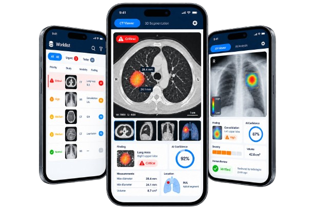
A DICOM-native radiology assistant designed to identify critical findings within seconds. Using a medical foundation model and 3D volumetric analysis, the platform detects, segments, and quantifies multiple conditions from a single study. It integrates with PACS workflows, prioritizes urgent cases, explains its findings through visual heatmaps, and protects patient privacy through federated learning. The system supports radiologists in clinical decision-making without replacing their final judgment.
Project Duration
Not specified
Technology Used
Python, PyTorch, MONAI, nnU-Net, Vision Transformers, 3D CNNs, MAE, DINO, Grad-CAM, pydicom, NVFlare, Flower, ONNX, TensorRT, PACS, HL7 FHIR, MLflow.
Country
Global
Problem
Increasing scan volumes were placing pressure on limited radiology teams.
Critical findings could remain unread in long worklists.
Most AI tools evaluated only one medical condition at a time.
Classification-only systems could not locate or measure abnormalities.
Black-box predictions were difficult for radiologists to verify.
Patient data could not be transferred outside hospitals without privacy and regulatory risks.
Solutions
Developed one foundation model capable of analyzing multiple conditions.
Used 3D volumetric models to detect, segment, and quantify abnormalities.
Integrated the system with DICOM and PACS workflows.
Added calibrated confidence scores and uncertainty alerts.
Generated visual heatmaps to help radiologists verify findings.
Automatically moved critical studies higher in the worklist.
Used federated learning to train models without moving patient data outside hospitals.

Project Workflow
Research
Competitors Analysis
Concept Ideation
Experimentation and optimization
Visual Design
Development
Testing
Results

Critical findings can be flagged within seconds of scan acquisition.

Urgent studies are prioritized ahead of routine cases.

Detection and measurement become more consistent across studies.

Low-confidence findings are automatically referred for human review.